I'm a huge fan of the Khan Academy and regularly watch his videos when I have a question about something in mathematics. Usually it only takes five or so minutes into the video for me to recall how to accomplish the task, and I can move along. I've always wanted to see if I could do the same thing for ECG interpretation.
So here is my inaugural attempt; rapid axis determination using leads I and aVF (assuming you're ok with a ±5° difference):
Monday, 23 May 2011

Wednesday, 6 April 2011
Conclusion to 54 year old female CC: BLS intercept
As many readers noted, there is a lot of baseline wander. This is not the most helpful of 12-Leads. On scene the crew attempted multiple 12-Leads, however, the patient would not sit still and that was the best one.
I think a close look at the Initial 12-Lead has enough information to make a field diagnosis.
Tuesday, 5 April 2011
New Case Study at EMS 12-Lead Blog
My first case study as an associate editor is up at the EMS 12-Lead Blog, so check it out: 54 year old female cc: BLS Intercept:
Enjoy.
Also, I've done a brief review of atrioventricular blocks to help with identification of the rhythm in this case study!"It is just after 3am when you are called to intercept a BLS unit on scene with a 54 year old female with a low heart rate.Upon your arrival, you find two EMT-Basics attending to a small woman lying in bed, who appears acutely ill..."
Enjoy.
Thursday, 10 March 2011
Unrecognized Limb Lead Misplacement?
Dr. Smith's ECG Blog has a new case up, "Reperfusion through collaterals associated with nitroglycerin, lateral MI with reciprocal T-wave inversion in lead III," with a pretty stark change in the initial 12-Leads. However, I have a hunch the stark change was really a change in the limb lead positions!
Look at leads I and II, notice how they "swap" positions between the two 12-Leads. Now look at aVL and aVF, notice how the "swap" positions too. Now take a look at lead III. It goes from inverted P's and T's with a Qr complex, to upright P's and T's with a Rs complex.
I propose that this change is due to a simple reversal of two leads. If we take a look at our friend Einthoven's Triangle (we covered this in a previous post on the S5 Lead) we can see that this makes sense!
We can see that Lead I is actually looking at Lead II and Lead II is actually looking at Lead I; confirmed with ECG's 1 and 2. Lead III becomes an inverted Lead III; confirmed again in the original ECG's. This looks like a case of an unrecognized left arm and left leg lead reversal.
What I find most interesting is if you compare every ECG except the first, it appears to be a case with subtle posteriolateral changes that may have been missed had there not been the lead reversal!
 |
| ECG 1 and ECG 2: Limb Leads Only |
I propose that this change is due to a simple reversal of two leads. If we take a look at our friend Einthoven's Triangle (we covered this in a previous post on the S5 Lead) we can see that this makes sense!
 |
| LA/LL Swap: Einthoven's Triangle is "Flipped" |
What I find most interesting is if you compare every ECG except the first, it appears to be a case with subtle posteriolateral changes that may have been missed had there not been the lead reversal!
Monday, 28 February 2011
Highlighting Atrial Activity on an ECG: The S5 Lead
Kelly Grayson, of A Day in the Life of an Ambulance Driver fame, posted an article on EMS1.com over a year ago entitled The Leads Less Traveled. In this he touched on modified chest leads (MCL1 through MCL6), right precordial leads (V4R), and the S5 Lead.
Update: after posting this I have since learned it is also known as the Lewis Lead, after Sir Thomas Lewis1, and have included a link to an article detailing how it was derived.
I had never heard of the S5 Lead before and promptly forgot about it until yesterday, when I finished acquiring 12-Leads for my limb lead reversal project. I went ahead and captured a rhythm strip from myself using the S5 lead placement.
Before we cover the S5 Leads, let's recap normal lead placement and our friend, Einthoven's Triangle. This produces convenient ECGs with positive waveforms along the usual mean vector of the heart. Lead I points to 0°, Lead II points to 60°, and Lead III points to 120°.
Additionally, the electrodes themselves are placed out on the limbs which generally results in waveforms proportional to the myocardium involved. Atrial activity is shown as well, but considering the proportion of myocardium involved in atrial depolarization, this configuration is not always useful in finding P-waves.
Now let's introduce the S5 Lead. You can produce this using many variations of the electrodes, however, for simplicity's sake we will stick with Kelly's description:
Update: after posting this I have since learned it is also known as the Lewis Lead, after Sir Thomas Lewis1, and have included a link to an article detailing how it was derived.
I had never heard of the S5 Lead before and promptly forgot about it until yesterday, when I finished acquiring 12-Leads for my limb lead reversal project. I went ahead and captured a rhythm strip from myself using the S5 lead placement.
 |
| S5 Leads: monitoring Leads I and II. |
 |
| Our friend, Einthoven's Triangle. |
 |
| Normal Placement: Leads I and II from the same patient. |
- Place the Right Arm electrode on the patient's manubrium.
- Place the Left Arm electrode on the 5th intercostal space, right sternal border.
- Place the Left Leg electrode on the right lower costal margin.
- Monitor Lead I.
 |
| Maximal atrial activity monitoring Lead I, S5 Lead configuration. |
Notice the change in the direction of each lead. Lead I now points to the usual mean vector of atrial depolarization. Lead II and lead III are nearly perpendicular to the usual mean vector of ventricular depolarization. What does this mean for the electrocardiographer? If you remember that a vector which travels towards a lead is positive and perpendicular to a lead is isoelectric the answer is easy: atrial activity is highlighted, ventricular activity is diminished.
 |
| S5: Lead I |
 |
| S5: Lead II |
The new direction Lead I points in is not quite perpendicular to the mean vector and it is also closer to the ventricular depolarization, hence we still have clear ventricular activity. However, the direction and location of Lead I is right in front of the atrial depolarization wavefront, giving clear P-waves. Lead II shows a large P-wave and small, nearly isoelectric ventricular activity.
If I can remember, I will try and acquire S5 Leads in the field. Has anyone else used the S5 Lead? Are there any other interesting lead configurations we should use?
- Bakker, ALM, et al. The Lewis Lead: Making Recognition of P Waves Easy During Wide QRS Complex Tachycardia. Circ (2009); 119:e592-e593. [Free Full Text]
Monday, 14 February 2011
Limb Lead Reversal: Preliminary Findings
Back in January, Tom over at the EMS 12-Lead blog had an interesting case entitled "Bait and Switch" in which the diagnosis of a STEMI was potentially masked due to incorrect limb lead placement. Interestingly, the limb lead placement was not one of classic LA/RA reversal, but rather a "rotation" of the limb leads. In this instance, the cardiac monitor did not detect the incorrect limb lead positioning. Over the last few weeks I have set out to collect 12-Lead ECGs acquired from each of the 24 possible limb lead positions and to catalog the characteristics of each.
All of the ECGs I have acquired are on LifePak 12 monitors using the GE Marquette 12SL algorithm. Currently, only classic limb lead reversal has produced the, "*** Suspect arm lead reversal, interpretation assumes no reversal," message. However, I still have 12 combinations of lead placements to complete.
Here are 3 ECG's acquired from a healthy male subject without any known cardiac abnormality or history (i.e. me).
All of the ECGs I have acquired are on LifePak 12 monitors using the GE Marquette 12SL algorithm. Currently, only classic limb lead reversal has produced the, "*** Suspect arm lead reversal, interpretation assumes no reversal," message. However, I still have 12 combinations of lead placements to complete.
Here are 3 ECG's acquired from a healthy male subject without any known cardiac abnormality or history (i.e. me).
 |
| Normal ECG **Unconfirmed**; Normal Sinus Rhythm |
 |
| Abnormal ECG **Unconfirmed**; *** Suspect arm lead reversal, interpretation assumes no reversal; Normal sinus rhythm; Right axis deviation; Nonspecific ST abnormality. |
 |
| Abnormal ECG **Unconfirmed**; Unusual P-axis, possible ectopic atrial rhythm; Left axis deviation; ST & T wave abnormality, consider inferior ischemia |
Tuesday, 21 December 2010
Harvard's ECG Wave Maven
I am constantly searching for resources which let me hone my electrocardiography skills and would like to share a gem I discovered a few months ago. Harvard's School of Medicine and the Beth Israel Deaconess Medical Center has an excellent resource: ECG Wave Maven: Self-Assessment Program for Students and Clinicians. You can browse their cases as a quiz or for reference, and each case includes high resolution ECGs for your inspection.
I've found their difficulty ratings to be pretty accurate, and I've found that Level 3 or less (of 5 difficulty levels) are all ECG findings that Paramedics should be able to recognize.
I encourage all of you to go spend a few hoursdays at the site brushing up on your ECG interpretation skills, your patients deserve it!
I've found their difficulty ratings to be pretty accurate, and I've found that Level 3 or less (of 5 difficulty levels) are all ECG findings that Paramedics should be able to recognize.
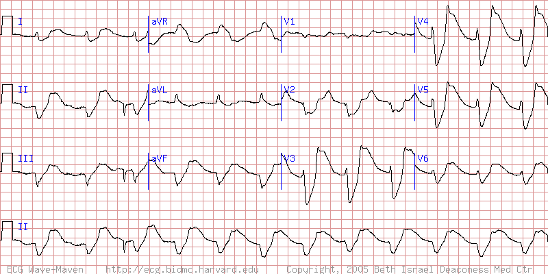 |
| Nathanson L A, McClennen S, Safran C, Goldberger AL. ECG Wave-Maven: Self-Assessment Program for Students and Clinicians. http://ecg.bidmc.harvard.edu. |
Subscribe to:
Posts (Atom)